Osteotomy literally means cutting of bone. During an osteotomy the bone is cut and it’s position is changed to a new position. This is done to shift the load from one part of a joint to another or to treat instability of the knee from ligament damage.
In some people the inside compartment of the knee wears out faster than the outside. This is called medial compartment arthritis. The outside or lateral compartment remains relatively normal and not badly affected by arthritis. The idea of the operation is move the weight from the damaged medial compartment to the more normal lateral compartment. This relieves the pain and yet leaves the person with their own knee rather than having a knee replacement (more about this later).
In certain patients with anterior cruciate ligament (ACL) tears just reconstructing the ligament is not enough. These patients may require an HTO or an HTO combined with an ACL reconstruction.
We know that osteotomy only works for some patients. The patients most suitable for a high tibial osteotomy are:
- Active
- Generally young (under 55 years of age)
- Have arthritis affecting one part of the joint
- Have a good range of motion of the joint
- Have good bone quality
- Able to use crutches easily
- Committed to rehabilitation
Certain groups of patients are not suitable for a high tibial osteotomy:
To watch a video animation of an osteotomy click here
- Inflammatory arthritis
- Infection
- More than one compartment involved
- Stiffness
- Obesity
- Over 65 years of age
What Is Done During an Osteotomy operation?
Each osteotomy is individualised to the needs of the patient. Special Xrays are taken called long leg (or 3ft) weight bearing xrays of the leg. This allows me to measure the weight bearing axis of your leg (to see where the load falls in your knee). I can then calculate exactly how far to shift the bone to allow the weight to fall through the correct part of the knee. Careful planning before the surgery is vital to the success of the operation.
While it is possible to perform several different types of osteotomies the medial opening wedge osteotomy is the one that is most commonly used: All osteotomies need to be held in place with some form of device, usually a plate and screws. If the correction is very large a bone graft may be needed but this is not very common.
- Closing Wedge – where a triangular piece of bone is cut out,
- Opening Wedge – where the bone is spread open, and,
- Dome Osteotomies – where the bone is cut in a semicircular shape.
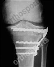

The operation is performed under general anaesthetic. A cut is made over the medial or inside part of the upper tibia (shin bone) and a wire is drilled into the bone to establish the correct position for the bone cut. An xray is taken to confirm that the wire is in the correct position and then the bone is cut with a saw and an osteotome (chisel). Once the appropriate correction has been achieved a plate and screws are inserted to hold the bone in that position.
A brace is applied to the leg which is used for at least 6 weeks. The brace allows movement of the knee and is removed for showers. This is a huge advance from the days when a plaster cast was applied for 6 weeks after this operation.
You will stay overnight in hospital and get up the next day using crutches. Most people go home the day after the operation. Your operated leg will need to remain off the ground (Non Weight Bearing) for 6 weeks after the surgery. After that you are allowed to take some of your weight on the leg for 6 weeks and full or unrestricted weight bearing is allowed at 3 months if the xray shows good bone healing. Most people find it more comfortable to wear their brace for the full 3 months but it can be removed after 6 weeks if needed. The brace can be worn under or over clothing and is not heavy or cumbersome.
Once the bone has healed there are no restrictions on what you can do! This is the main advantage of an osteotomy. You do not have metal or plastic parts replacing your knee. Many people are able to return to vigorous activities such as team sports and are able to continue to work in labouring or heavy manual jobs. Very few people return to running or jogging.
If you work sitting at a desk you should be able to return to work 10-14 days after the operation. If you need to stand or walk a lot or lift heavy objects then 3 months or more may be required off work.
In the right patient an HTO is an excellent operation but tibial osteotomy does not give the same quality of symptom relief from arthritis that is usually seen with total knee replacement. It does not reverse arthritis but does stop its rapid progression. A few patients (5-8%) are not improved and 2% are worse off. In general, 60% – 75% of osteotomies will still be functioning very well ten years later. This means a knee relatively free of pain with high level function possible.
The others will have had progression of their arthritis and need a total knee replacement. Unfortunately the results of a total knee replacement after an HTO are not quite as good as if the knee had never had surgery but you will have had many years of extra usage of the knee without restriction. It is considerably easier and achieves better results than performing a revision total knee replacement (redoing a knee replacement once it has worn out). This is usually a good trade off for active people and remains a worthwhile operation for these people.